Characterization of Biomaterials 227
at this critical length scale. It has been used to study the hierarchical structure of a
biological material such as a bone. In particular it plays a crucial role in studying
the lowest hierarchical level, within the nanometer scale, where the distribution
of organic fibers (collagen) and crystalline mineral nano-particles has a major
influence for mechanical optimization of bone.
4
10.3 SURFACE ANALYSIS METHODS
The surface structure characterization of a biomaterial presents different analyt-
ical challenges since the composition and spacial arrangement of the molecular
segments on the surface may not be representative of the bulk structure. In
addition, the surface is the interface where the biomaterial meets and interacts
with the molecular constituents of the biological host setting (e.g., bone, soft tissue,
blood, cells, body fluids). These interactions can modify the material and result in
macroscopic events in the biological media which ultimately determine whether
or not an MEMS device will have successful performance in the host biological
environment. Therefore, surface characterization will be discussed in more detail
in this chapter.
The properties that are of interest in the characterization of biomaterial sur-
faces include the chemical structure, the hydrophilicity or hydrophobicity, the
presence of ionic groups, the morphology and topography.
1,3
Varying degrees of
information about these properties can be obtained using different analysis meth-
ods including microscopic, spectroscopic and thermodynamic and other methods.
We aim to provide an overview on some of the specific techniques frequently used
in the characterization of biomaterial surfaces. In addition, discussions and refer-
ences are made to emerging methods such as rapid Raman spectroscopy, optical
coherence tomography (OCT), and multi-photon excitation microscopy that have
recently come to the forefront of biomedical research and possess great potentials
for studying the interactions of biomaterials with real biological environment
in vivo.
10.3.1 Microscopic Methods
Microscopy techniques often used in biomaterial analysis include scanning elec-
tron microscopy (SEM), transmission electron spectroscopy (TEM), scanning tun-
neling microscopy (STM) and atomic force microscopy (AFM).
11
Recent develop-
ments in light microscopy include confocal laser scanning microscopy (CLSM) and
optical near-field microscopy.
12
10.3.1.1 Electron Microscopy
An electron microscope is a type of microscope that uses electrons to illuminate
a specimen in a vacuum environment and create an enlarged image. Electron
microscopes have much greater resolving power than light microscopes. Electron
SO13997_text.indd 235SO13997_text.indd 235 26/01/2011 3:51 PM26/01/2011 3:51 PM
228 H. Zeng
microscopy can be performed in either transmission or secondary electron modes.
In transmission electron microscopy (TEM),
13
images are formed by diffraction
or phase contrast mechanisms. The detectors are phosphor screens coupled
with digital CCD cameras. Unlike the TEM, where electrons of the high energy
beam form the image of the specimen, the scanning electron microscope (SEM)
14
produces images by detecting low energy secondary electrons which are emitted
from the surface of the specimen due to excitation by the primary electron beam. In
the SEM, the electron beam is rastered across the sample, with detectors building
up an image by mapping the detected signals with beam positions. SEM and TEM
are commonly used for studying both the surface morphology of and the cellular
response to biomaterials.
15,16
SEM sample preparation involves fixation (when proteins, cells, or tissues
are present), followed by drying, attachment to a metallic stub, and then coating
with a metal prior to data collection. The thin metallic coating is typically 20 to
30 nm in thickness. Common conductive metals used include gold, platinum,
or gold/palladium alloy. It should be noted that the drying and metal coating
processes used in the preparation of some polymeric materials might alter surface
morphology, particularly those surfaces that may undergo changes in a hydrated
environment. In addition to imaging the surface morphology of biomaterials, the
SEM can be combined with other analysis methods such as energy dispersive X-
ray analysis to determine elemental distribution
15
and IR and Raman spectroscopy
to monitor surface modification procedures.
17
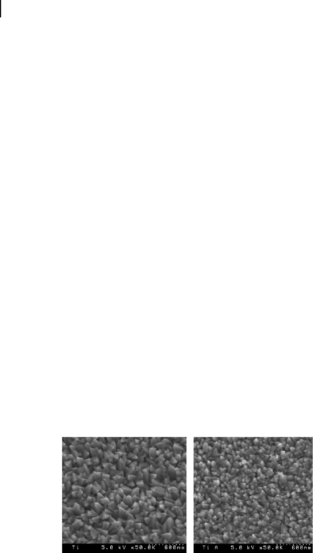
Figure 10.2 shows an example
application of using SEM to assess the effect of sterilization on biomaterial surface
morphology.
18
TEM sample preparation involves fixation (if biological materials are present),
processing, embedding and sectioning. Samples must be very, very thin (50 nm–
90 nm). This is achieved by embedding samples in hard plastic resins and
using very sharp diamond knife ultramicrotomes. TEM provides extraordinary
resolution of organelle and membrane structures in single cells. There is probably
Figure 10.2. SEM micrograph of as-deposited surface finish of sputter deposition elemen-
tal titanium (left side) compared with SEM micrograph of surface finish of autoclaved
sputter deposition elemental titanium (right side). Pictures were taken from a candidate
biomaterial for developing implantable MEMS devices. With permission from.
18
SO13997_text.indd 236SO13997_text.indd 236 26/01/2011 3:51 PM26/01/2011 3:51 PM
Get Biomaterials for MEMS now with the O’Reilly learning platform.
O’Reilly members experience books, live events, courses curated by job role, and more from O’Reilly and nearly 200 top publishers.

