Chapter 1Defining a Science Image
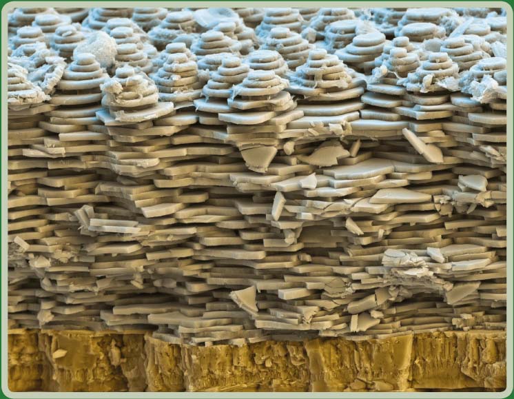
Science or art? This scanning electron photomicrograph features the shell of the white-lipped snail, Cepaea hortensis. The shell is composed of lime. The photograph reveals the layers of the shell, including where the inner wall forms new crystal platelets, and is graphically very interesting. This image is a composite from three detectors, one secondary detector and two backscattered electron-detectors. Compositingand coloring was accomplished using Adobe Photoshop software. The scanning electron microscope used 10 kV with a 13 mm working distance and 2500:1 at 15 ×13 cm. The shell was photographed ...
Get Laboratory Imaging & Photography now with the O’Reilly learning platform.
O’Reilly members experience books, live events, courses curated by job role, and more from O’Reilly and nearly 200 top publishers.

