April 2019
Intermediate to advanced
233 pages
6h 42m
English
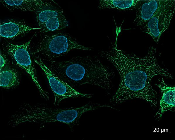
In this chapter, we introduce you to deep learning techniques for microscopy. In such applications, we seek to understand the biological structure of a microscopic image. For example, we might be interested in counting the number of cells of a particular type in a given image, or we might seek to identify particular organelles. Microscopy is one of the most fundamental tools for the life sciences, and advances in microscopy have greatly advanced human science. Seeing is believing even for skeptical scientists, and being able to visually inspect biological entities such as cells builds an intuitive understanding of the underlying mechanisms of life. A vibrant visualization of cell nuclei and cytoskeletons (as in Figure 7-1) builds a much deeper understanding than a dry discussion in a textbook.
The question remains how deep learning can make a difference in microscopy. Until recently, the only way to analyze microscopy images was to have humans (often graduate students or research associates) manually inspect these images for useful patterns. More recently, tools such as CellProfiler have made it possible for biologists to automatically assemble pipelines for handling ...
Read now
Unlock full access


.jpg)