Skip to Content
Laboratory Imaging & Photography
book

Laboratory Imaging & Photography

by Michael Peres
January 2017
Intermediate to advanced
392 pages
15h 38m
English
Routledge
Content preview from Laboratory Imaging & Photography

Chapter 9Light Microscopy


fig9_1

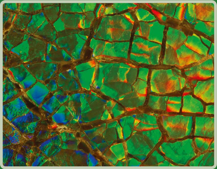
This photograph features the shell of a fossilized ammonite that was hundreds of millions of years old. The iridescent colors are produced by alternating layers of aragonite and conchiolin, or from minerals that have replaced them over the millions of years during the fossilization process. The shell was photographed with a simple microscope. Axial illumination was used to show the iridescent colors. The specimen was coated with mineral oil to control specular highlights. Creating effective focus at high magnifications is always difficult. This photo was originally published in Ancient Microworlds, published ...

Become an O’Reilly member and get unlimited access to this title plus top books and audiobooks from O’Reilly and nearly 200 top publishers, thousands of courses curated by job role, 150+ live events each month,
and much more.
Start your free trial

You might also like

Digital Ultraviolet and Infrared Photography

Digital Ultraviolet and Infrared Photography

Adrian Davies
Microscope Image Processing

Microscope Image Processing

Qiang Wu, Fatima Merchant, Kenneth Castleman

Publisher Resources

ISBN: 9781317593003