December 2007
Intermediate to advanced
224 pages
7h 18m
English
This section briefly describes the most important parts of the human auditory system. For more detailed information the reader is referred to dedicated textbooks, e.g. [226].
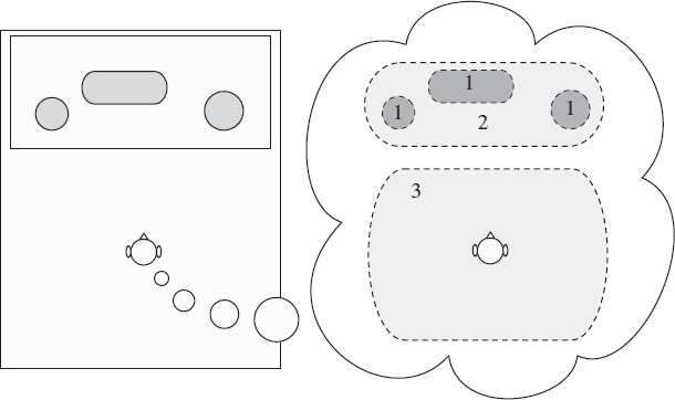
Figure 3.1 Illustration of the perception of the auditory spatial image. Perception of auditory objects (1); extent of the auditory spatial image (2); and listener envelopment (3).
Changes in the acoustical pressure are received by the outer ear, which consists of a visible part (the pinna) and a canal leading to the eardrum (the tympanic membrane). Attached to the eardrum are the middle ear bones (ossicles): the malleus (the outermost), the incus and the stapes. The foot of the stapes is connected to the oval window, which is part of the inner ear. The major function of the middle ear is to ensure the efficient transfer of sound from the air to the fluids of the cochlea. It acts as an impedance-matching device that improves sound transmission.
The cochlea within the inner ear is composed of a bony labyrinth. It is partitioned into three fluid-filled tubes. These tubes are separated by membranes: between the first (scala vestibuli) and second (scala media) there is Reissner's membrane. The scala media is in turn separated from the lower space (scala tympani) by the basilar membrane.
The so-called organ of Corti spirals on the basilar ...
Read now
Unlock full access


