32.6 Real MR Brain Image Experiments
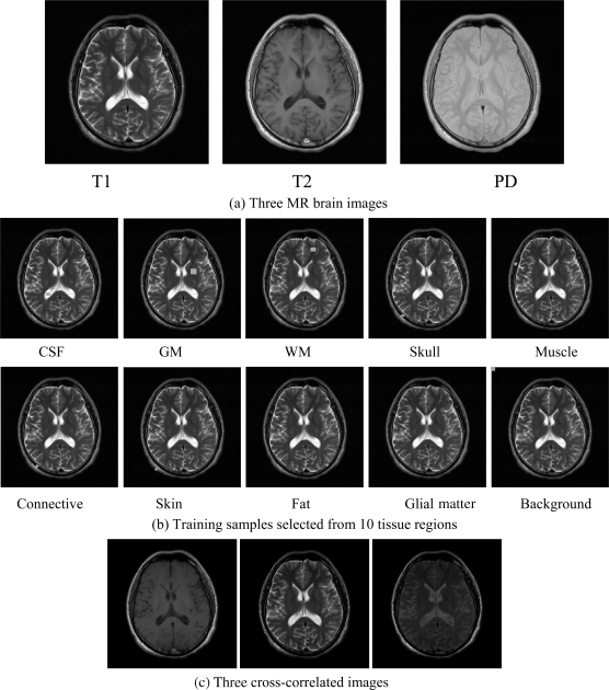
The real MR images used for experiments were obtained in the TaiChung Veterans General Hospital (TCVGH) to further validate the utility of the proposed LSMA-based intrapixel techniques in real images in calculating partial volumes of brain tissues. The MR images shown in Figure 32.23(a) were obtained from one normal volunteer by a whole body 1.5-T MR system (Sonata, Siemens, Erlangen, Germany). The routine brain MR protocol consisted of axial spin echo T1 weighted images (TR/TE = 400/9 ms), proton density image (TR/TE = 4000/10 ms), and T2 weighted images (TR/TE = 4000/91 ms). Other imaging parameters included for this experiment were slice thickness = 6 mm, matrix = 256 × 256, field of view (FOV) = 24 cm, and number of excitations (NEX) = 2. These images are shown in Figure 32.23(a). Training sample sets for 10 classes were selected according to their anatomical structures shown in Figure 32.23(b).
Figure 32.23 Three real MR images in (a) along with training samples in (b).

By visual inspection of Figures 32.23–32.26, the PVE results produced by all the techniques separated the three major substances clearly in general. However, there were also some supposedly WM substances showing in GM-generated images by all the techniques. Moreover, similar to the synthetic brain image experiment, OSP and LSOSP produced similar estimated abundances and ...
Get Hyperspectral Data Processing: Algorithm Design and Analysis now with the O’Reilly learning platform.
O’Reilly members experience books, live events, courses curated by job role, and more from O’Reilly and nearly 200 top publishers.

