Think of the central nervous system like a mushroom with the spinal cord as the stalk and the brain as the cap. Most of the hacks in this book arise from features in the cortex, the highly interconnected cells that make a thin layer over the brain...but not all. So let’s start outside the brain itself and work back in.
Senses and muscles all over the body are connected to nerves, bundles of neurons that carry signals back and forth. Neurons come in many types, but they’re basically the same wherever they’re found in the body; they carry electric current and can act as relays, passing on information from one neuron to the next. That’s how information is carried from the sensory surface of the skin, as electric signals, and also how muscles are told to move, by information going the other way.
Nerves at this point run to the spinal cord two by two. One of each pair of nerves is for receptors (a sense of touch for instance) and one for effectors— these trigger actions in muscles and glands. At the spinal cord, there’s no real intelligence yet but already some decision-making—such as the withdrawal reflex—occurs. Urgent signals, like a strong sense of heat, can trigger an effector response (such as moving a muscle) before that signal even reaches the brain.
The spinal cord acts as a conduit for nerve impulses up and down the body: sensory impulses travel up to the brain, and the motor areas of the brain send signals back down again. Inside the cord, the signals converge into 31 pairs of nerves (sensory and motor again), and eventually, at the top of the neck, these meet the brain.
At about the level of your mouth, right in the center of your head, the bundles of neurons in the spinal cord meet the brain proper. This tip of the spinal cord, called the brain stem, continues like a thick carrot up to the direct center of your brain, at about the same height as your eyes.
This, with some other central regions, is known as the hindbrain. Working outward from the brain stem, the other large parts of the brain are the cerebellum, which runs behind the soft area you can feel at the lower back of your head, and the forebrain, which is almost all the rest and includes the cortex.
Hindbrain activities are mostly automatic: breathing, the heartbeat, and the regulation of the blood supply.
The cerebellum is old brain—almost as if it were evolution’s first go at performing higher-brain functions, coordinating the senses and movement. It plays an important role in learning and also in motor control: removing the cerebellum produces characteristic jerky movements. The cerebellum takes input from the eyes and ears, as well as the balance system, and sends motor signals to the brain stem.
Sitting atop the hindbrain is the midbrain, which is small in humans but much larger in animals like bats. For bats, this corresponds to a relay station for auditory information—bats make extensive use of their ears. For us, the midbrain acts as a connection layer, penetrating deep into the forebrain (where our higher-level functions are) and connecting back to the brain stem. It acts partially to control movement, linking parts of the higher brain to motor neurons and partially as a hub for some of the nerves that don’t travel up the spinal cord but instead come directly into the brain: eye movement is one such function.
Now we’re almost at the end of our journey. The forebrain, also known as the cerebrum, is the bulbous mass divided into two great hemispheres—it’s the distinctive image of the brain that we all know. Buried in the cerebrum, right in the middle where it surrounds the tip of the brain stem and midbrain, there’s the limbic system and other primitive systems. The limbic system is involved in essential and automatic responses like emotions, and includes the very tip of the temporal cortex, the hippocampus and the amygdala, and, by some reckonings, the hypothalamus. In some animals, like reptiles, this is all there is of the forebrain. For them, it’s a sophisticated olfactory system: smell is analyzed here, and behavioral responses like feeding and fighting are triggered.
For us humans, the limbic system has been repurposed. It still deals with smell, but the hippocampus, for example—one part of the system—is now heavily involved in long-term memory and learning. And there are still routing systems that take sensory input (from everywhere but the nose, which is routed directly to the limbic system), and distribute it all over the forebrain. Signals can come in from the rest of the cerebrum and activate or modulate limbic system processing common to all animals—things like emotional arousal. The difference, for us humans, is that the rest of the cerebrum is so large. The cap of the mushroom consists of four large lobes on each hemisphere, visible when you look at the picture of the brain. Taken together, they make up 90% of the weight of the brain. And spread like a folded blanket over the whole of it is the layer of massively interconnected neurons that is the cerebral cortex, and if any development can be said to be responsible for the distinctiveness of humanity, this is it. For more on what functions the cerebral cortex performs, read [Hack #8].
As an orienting guide, it’s useful to have a little of the jargon as well as the map of the central nervous system. Described earlier are the regions of the brain based mainly on how they grow and what the brain looks like. There are also functional descriptions, like the visual system [[Hack #13]], that cross all these regions. They’re mainly self-explanatory, as long as you remember that functions tend to be both regions in the brain and pathways that connect areas together.
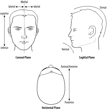
There are also positional descriptions, which describe the brain geographically and seem confusing on first encounter. They’re often used, so it’s handy to have a crib, as shown in Figure 1-1.
These terms are used to describe direction within the brain and prefix the Latin names of the particular region they’re used with (e.g., posterior occipital cortex means the back of the occipital cortex).
Unfortunately, a number of different schemes are used to name the subsections of the brain, and they don’t always agree on where the boundaries of the different regions are. Analogous regions in different species may have different names. Different subdisciplines use different schemes and conventions too. A neuropsychologist might say “Broca’s areas,” while a neuroanatomist might say “Brodman areas 44, 45, and 46”—but they are both referring to the same thing. “Cortex” is also “neocortex” is also “cerebrum.” The analogous area in the rat is the forebrain. You get the picture. Add to this the fact that many regions have subdivisions (the somatosensory cortex is in the parietal lobe, which is in the neocortex, for example) and some subdivisions can be put by different people in different supercategories, and it can get very confusing.
Three excellent online resources for exploring neuroanatomy are Brain Info ( http://www.med.harvard.edu/AANLIB/home.html ), The Navigable Atlas of the Human Brain ( http://www.msu.edu/~brains/humanatlas ), and The Whole Brain Atlas ( http://braininfo.rprc.washington.edu ).
The Brain Museum ( http://brainmuseum.org ) houses lots of beautifully taken pictures of the brains from more than 175 different species.
BrainVoyager ( http://www.brainvoyager.com ), which makes software for processing fMRI data, is kind enough to provide a free program that lets you explore the brain in 3D.
Nolte, J. (1999). The Human Brain: An Introduction to Its Functional Anatomy.
Crossman, A. R., & Neary, D. (2000). Neuroanatomy: An Illustrated Colour Text.
Get Mind Hacks now with the O’Reilly learning platform.
O’Reilly members experience books, live events, courses curated by job role, and more from O’Reilly and nearly 200 top publishers.


